Equipment;
-Scalpel
-Board
-Scissors
-Lab coat
-Chopping board
-Gloves
-Scalpel
-Board
-Scissors
-Lab coat
-Chopping board
-Gloves
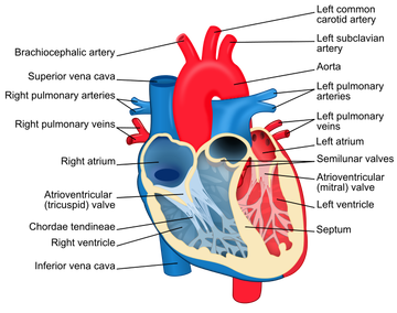 | 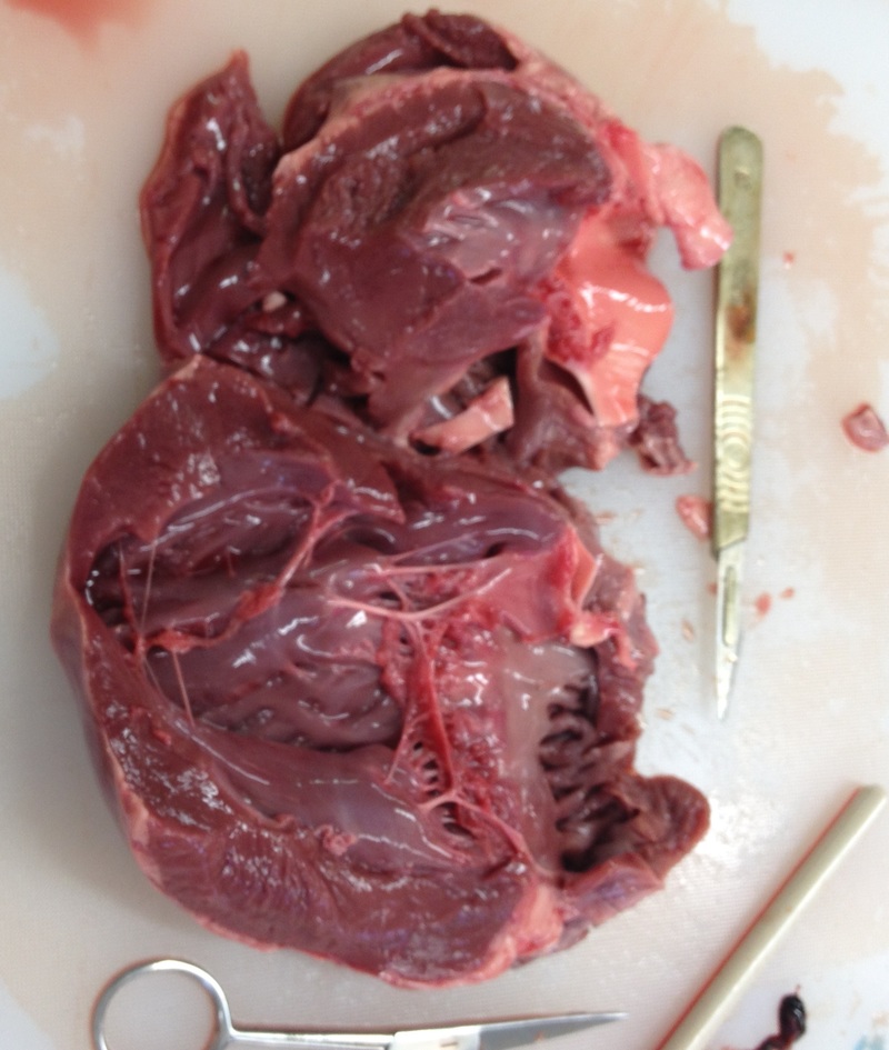 |
When comparing the heart diagram and the picture of the dissected heart, you are able to see the different sections. The section of the heart towards the bottom is the left ventricle, as it has the far thicker muscle as the blood has to be pumped across the body from that chamber. To the right of the dissected heart you can see the bicuspid valve, and further right is the left atrium. At the top of the right ventricle is the septum which separates the the left side of the heart from the right side.On the right side of the heart there is a far thinner muscular wall, as blood only needs to be pumped to the lungs which is close to the heart. At the right of this it is possible to see the lighter coloured muscle, this is the pulmonary artery, which carries deoxygenated blood to the lungs. The lighter muscle further up the picture is the vena cava, which carries the blood to the right atrium. Throughout the heart you can see the chordae tendineae (tendinous chords) which contract and relax to control the valves within the heart so that blood does not flow backwards.
Heart diagram;
https://en.wikipedia.org/wiki/File:Heart_diagram-en.svg
Heart diagram;
https://en.wikipedia.org/wiki/File:Heart_diagram-en.svg
 RSS Feed
